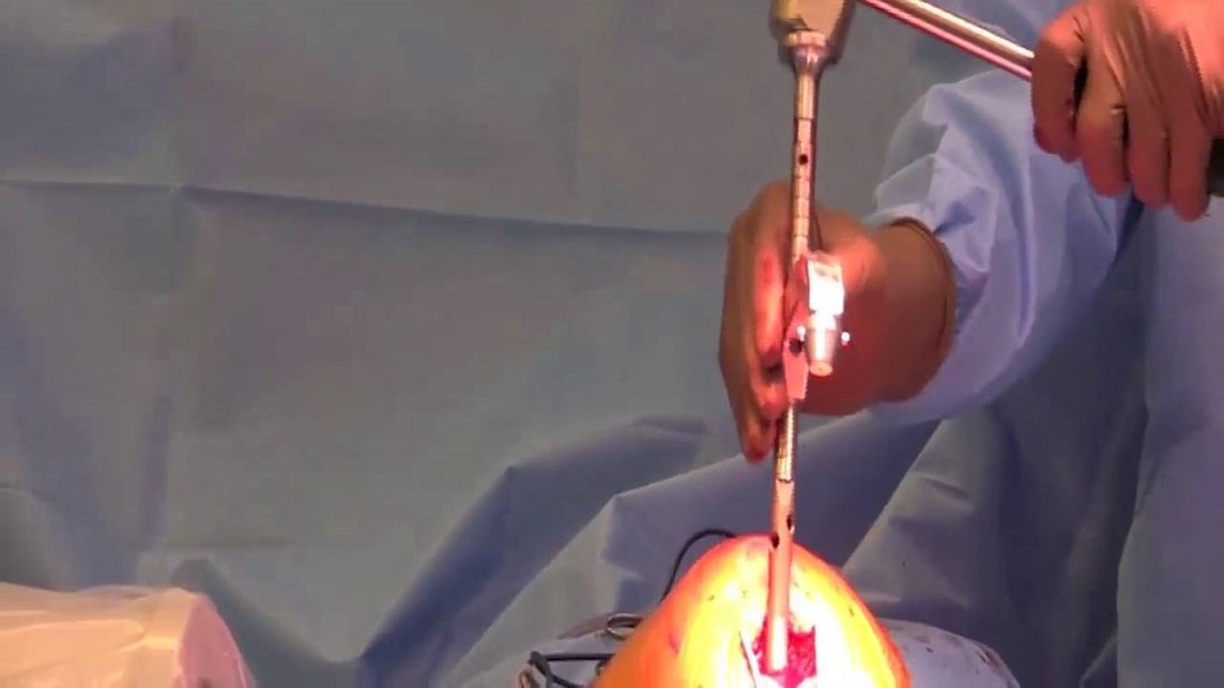
From the mechanical viewpoint, an orthopedic nail removal is not essential in a weight-bearing limb and dissimilar from a plate, it can be left indefinitely in the body. Removal initiated by the request of the patient should be delayed for eighteen months. Intramedullary devices sometimes induce local changes that can be irritative either to the bone or to the patient and require removal. Swelling and local pain secondary to backing out of the implant is another indication for removal; confirmed bone union on radiological examination is a prerequisite for such removal.
A sharp-angled deformation seeming in the follow-up roentgenogram is an indication of appliance failure. A sharply bent device is necessary to be removed and replaced as it has undertaken plastic deformation and is expected to fail with further weight-bearing. Bent nails may be removed forcefully straightening and extraction.
Nail removal shouldn’t be undertaken lightly. Specialized extraction equipment fitting the exact nail must be available. Although removal is often a straightforward process, mismatching equipment, nail breakage, damage to the threads in the proximal end of the nail and distortion in the bony anatomy preventing removal are common causes of difficulty.
Difficult situations in nail removal
- The extraction hook fails to grasp.
- Stack the canal round the hook with ball-tipped guide wires. Hold the bunch in a vice grip and hammer out to remove the nail.
- Bone growth along the track of the locking bone screws removed some time ago.
- Re-drill the holes with an appropriately large drill bit.
- The broken nail in a united fracture; the extraction hook fails to regulate the alignment as well as pulls the nail tight against the opposite cortex rather than out of the canal.
- Take away the proximal half of the nail. Pass a guidewire that is ball-tipped into the proximal canal till the ball is near the broken end of the nail. Ream the proximal canal up to the nail’s broken end to facilitate easy removal. Pass 2 or more guidewires into the distal nail fragment and impact them with a hammer. Grasp the bunch in a vice grip as well as hammer out to remove the nail.
- The broken solid nail in an ununited fracture.
- Take away the proximal half. Create a window in the bone distal to the tip of the nail as well as hammer the nail out. In the femur, arthrotomy of the knee joint can be needed. In the tibia, if the nail is short, make an opening in the medial malleolus to arrive till the nail.
- The protruding nail in a healed fracture and nail doesn’t move.
- Use specialized metal cutting equipment to remove the offending nail length.
- Impacted nail.
- Find the point of maximum hold under fluoroscopy. Create a longitudinal cut in the bone with the help of an oscillating saw or an osteotome at that point. Extend the cut either way till the bone springs open enough to allow removal of the nail. The method is suited best for a tibial nail and is employed only as a final resort.
Full weight-bearing may start immediately after orthopedic nail removal (intramedullary nail), but a coming back to vigorous sporting activity should be delayed until rehabilitation is attained.


