


Also known as the thigh bone, the femur is the longest and strongest bone in our body, and breaks in this bone are called femur fractures. The head of the femur connects the pelvic socket to form the hip joint. The strength of the femur plays an important role in providing the ability to stand and move. Not only this, but the bone also supports various muscles, tendons, and ligaments. A femur fracture is a break in this bone.
In this article, we will be discussing femur fractures in detail, followed by their causes, diagnosis, and treatment.
What is a Femur, and What Happens When it Breaks?
The femur, commonly known as the thigh bone, is the longest and strongest bone in the human body. Do you know where is femur bone is located? It connects the hip to the knee and supports much of the body’s weight during standing, walking, and running. Because of its strength and location, it typically requires a significant force, such as a car accident or a high-impact fall, to break.
When the femur breaks, it is considered a serious injury. Symptoms include intense pain, swelling, bruising, and the inability to bear weight on the affected leg. The leg may appear deformed or shorter than the other one. A broken femur often requires surgical intervention, usually involving the insertion of metal rods, plates, or screws to stabilize the bone. Recovery can take several months and typically involves physical therapy to regain strength and mobility. Left untreated, a femur fracture can lead to complications such as blood loss, infection, or improper bone healing.
What is a Femur Fracture? An Overview.
Despite being the strongest bone in our body, the femur is prone to breaking. The causes include high-intensity trauma like falls or automobile accidents. In most cases, the treatment requires surgery. After treatment, rehabilitation is required to regain strength and the range of motion of the injured area.
As a tremendous force is required for the femur to break, in the elderly above 60, femur fractures can occur because of low-energy trauma. Femur fractures are in the category of serious fractures. Let us see why.
Why is a Broken Femur a Serious Injury?
There are certain reasons why femur fractures are among serious fractures:
- Excessive blood loss may occur if there is an open fracture. This means the femur protrudes out by breaking the skin. Although all types of open fractures are severe.
- In some cases, the person may go into shock because of a femur fracture.
- There may be a risk of hip fracture, especially when the break is in the proximal femur. The risk of such cases is high in osteoporotic people.
- A break in the distal femur may increase the chances of knee fractures. This condition is common in people with osteoporosis or who have had knee replacement surgery.
What Are the Different Types of Femur Fractures?
Femoral fractures are classified based on the location and the pattern of fracture. Let us see them one by one.
Types of Femur Fractures based on the Location of Break
Femoral Head Fractures
The femoral head is the topmost part of the bone, which is also known as “the ball”. This part connects to the pelvic socket to form the hip joint. Thus, any break in this region is a femoral head fracture. The head of the femur is covered with cartilage, and this allows its smooth and frictionless movement. Femoral head fractures may also affect your hip and make it difficult/painful to move.
Femoral Neck Fractures
One of the commonest locations for fractures in the femur is the neck region. Along with pertrochanteric fractures, femur fractures account for around 90% of femoral fractures. The femoral neck is the region just below the head of the femur. Elderly adults with poor bone density are ideal candidates for experiencing femoral neck fractures. Now, based on the location of the break in the femoral neck, these fractures can be classified into three types:
- Fracture in the sub-capital region; it is the junction of the femoral head and neck
- Fracture in the transcervical region; it is the mid-portion of the femoral neck
- Fracture in the basicervical region; it is the base of the femoral neck
Femoral Shaft Fractures
High-energy traumas are among the commonest causes of femoral shaft fractures. Men are more prone to breaking the femoral shaft. This is when the break occurs in the shaft of the femur. Being highly vascularized bone, the fractures in the femoral shaft may accompany neurovascular injury, and it may occur because of the:
- High-energy trauma
- Pathological fractures
- Bisphosphonate-related fractures
- Fragility fractures that occur because of low-energy trauma in the elderly
Femoral Condyle Fractures
The femoral condyle is a ball-shaped projection at the distal femur. Two femoral condyles, i.e., medial and lateral condyles, are there in each leg. Hence, a break at this region of the femur is a femoral condyle fracture.
Types of Femur Fractures Based on the Severity and Pattern of Break
Stress Fractures
Overuse or repetitive use of the femur results in stress fractures. In these conditions, a tiny crack develops in the bone. Athletes engaged in high-impact sports are at high risk of developing stress fractures. Besides this, occupational activities that require physical labor also make people prone to stress fractures. These types of fractures may take more time than other fractures to show symptoms.
Severe Impaction Fractures
These are the types of fractures in which the ends of the bone are forced toward each other because of high-intensity trauma. These fractures are severe and may cause the bone to break into multiple pieces.
Incomplete Fractures
Also known as partial fractures, this is when the femur breaks but not all the way through. Here, the break may occur in one or more places.
Completely Displaced Fractures
This is when the bone breaks and the broken bone fragments significantly move away from the line of the bone. Such fractures require surgical intervention.
What Are the Causes of Femur Fractures?
As the femur is the strongest bone in the body, a strong force is a must to break it. Thus, the causes of femur fractures include high-intensity trauma like:
- Automobile accidents
- Falling from a height
- Contact sports injuries
- Direct impact on the bone
- Gunshots
In the elderly, low-impact trauma like falling in the bathroom or hitting with something while walking can also break the femur. Besides this, people suffering from osteoporosis may also break their femur because of minor injuries or without any significant cause.
What Are the Symptoms of Femur Fractures?
Fractures are always painful and being a serious condition, femur fractures result in severe pain. Along with this, other symptoms that may occur include:
- Inability to put weight on the injured leg
- Difficulty in standing or walking
- In case of open fractures, bone fragments may break out of the skin and bleeding occurs
- Bruising in the thigh
- Swelling in the thigh region
- The injured leg may appear shorter when compared to the uninjured leg
- The injured leg may turn out, away from the body
How do Healthcare Service Providers Diagnose Femur Fractures?
The diagnosis of a broken thighbone requires a thorough physical examination followed by imaging tests. The physical examination involves the assessment of visible signs and symptoms. Thus, the healthcare service provider will look for swelling in the thigh along with bruising. With this, he will also identify ant deformity in the leg. The orthopedic specialist may also ask the patient to move the leg to check the range of motion and ability to put weight on the injured leg. However, imaging tests confirm the fracture along with its severity:
X-ray Examination
X-ray reports will depict the break and its type in the femur. This will give a better idea to the orthopedic specialist about the condition.
CT scan
CT scans are best for diagnosis in severe cases to check damage to the surrounding structures like muscles, tendons, ligaments, and nerves. This test produces detailed images of the bone and supportive structures.
Magnetic Resonance Imaging (MRI)
MRI can show damage to blood vessels, infections, and affected blood supply if there. MRI is recommended when the orthopedic specialist is in doubt regarding the fracture and its severity. The test provides clear images of the non-bony parts of the injured area.
What is the Treatment for a Broken/Fractured Femur?
Well, the treatment depends on the severity of the fracture but, most cases require surgical intervention. Non-displaced minor breaks in the femur may be fixed by immobilizing the bone using a splint or a cast. Sometimes, the orthopedic specialist may initially apply a cast to improve symptoms and surgery is later performed to properly realign the broken bone.
The surgical options available for the treatment of femur fractures include:
- Open Reduction and Internal Fixation (ORIF)
- External Fixation
- Intramedullary Nailing
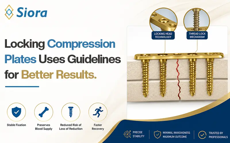
Open Reduction and Internal Fixation (ORIF)
The most common surgery to fix displaced femur fractures is ORIF. During the procedure, the orthopedic surgeon will make an incision at the hip or knee joint to access the fractured site. After this, the surgeon will remove damaged tissues, clean the area, and apply a metal orthopedic implant to stabilize the femur. The displaced fractured bone fragments are also brought back to their correct anatomy. Femur implants are used during surgical interventions.
External Fixation
Severe femur fractures like open fractures or comminuted fractures require external fixation. This is because such cases cannot be managed with ORIF. An external fixation system is the option for treatment in such cases. Here, the orthopedic surgeon will hold bone fragments using pins, orthopedic plates, rods, and bone screws and stabilize those implants to an external frame. This frame remains outside the body. In such conditions, a bone graft may also be used to compensate for the lost bone because of the fracture.
External fixation is also an option when other life-threatening injuries need to be addressed first. This is a quick and effective method to temporarily fix bones in the correct position.
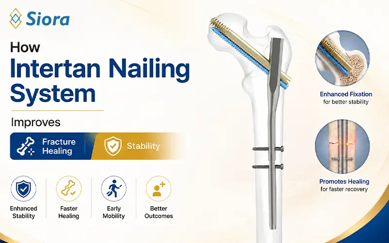
Intramedullary Nailing
When other treatment procedures are not feasible, intramedullary nailing is the choice of orthopedic surgeons. During the procedure, the surgeon inserts a special implant known as an intramedullary nail into the center of the femur. That region is known as the medullary cavity of the bone. This surgery allows the patient to put some amount of weight on the injured bone.
Note: All surgical procedures require local or general anesthesia.
What Recovery is Like After Femur Surgery?
The severity of the fracture decides how long it will take for the fracture to heal. Although young patients recover faster than the elderly.
For quick and improved recovery, it is important to follow post-surgical instructions given by the surgeon. They may include avoiding weight on the operated area for some time and using assistive devices to walk around, if necessary. It is also necessary to take care of the surgical wound as advised to prevent infection. For pain management and preventing infection, the doctor may prescribe analgesics and antibiotics.
Above all, rehabilitation is a must to get a previous-like range of motion and strength in the fractured leg. A physiotherapy specialist will help the patient get back to normal life as early as possible. Besides this, he will also guide you on how to safely perform routine tasks.
In the early stages of recovery, low-impact exercises are the best, along with weight-bearing ones. Remember, your seriousness and dedication during rehabilitation will play a vital role in deciding how fast you’ll recover.
Siora Surgicals Pvt. Ltd. is an experienced and one of the oldest orthopedic implants manufacturers in India. Operating for over 30 years, the company produces a CE-certified range of orthopedic implants and instruments, including external fixation systems, intramedullary nails, orthopedic implant plates, and more. We are also an experienced OEM/contract manufacturing service provider across the globe.